The Question No One Had Asked
How a 25 year-old technology transformed modern medical imaging

Nuclear magnetic resonance (NMR) is a measurement technique whose underlying physics was discovered by Columbia University professor Isidor Rabi in 1938, for which he won the Nobel Prize in Physics in 1944. Eight years later, two physicists, Felix Bloch and Edward Purcell, shared a second Nobel Prize for extending Rabi’s discovery to a practical application that worked for everyday materials. For the next 25 years, NMR played an indispensable role as a laboratory technique that helped physicists and chemists measure and thereby understand the fundamental nature of matter. By 1970, NMR equipment was standard in serious chemistry labs around the world — but the use was limited to determining the molecular structure of materials. And it was destined to remain there until an insight by Paul Lauterbur, a professor of chemistry and radiology at State University of New York at Stony Brook, who sketched out a fundamentally new concept over lunch with a colleague in September, 1971. His insight — that NMR signals could carry both chemical and spatial information, and thus could be turned into actual images — led to the development of new technologies that transformed medical imaging.
The setting of the lunch proved to be auspicious. In the summer of 1971, following the end of the semester at Stony Brook, Lauterbur had agreed to step in short-term as the unpaid interim leader of NMR Specialties, a western Pennsylvania company that sold and leased state-of-the-art NMR equipment, to try to save the company from bankruptcy. He had joined the board of directors some years prior, and the company’s bank was refusing further support unless a trusted person could lead the company out of the crisis. At the time, Lauterbur was a preeminent scientist in NMR research, with 20 years of experience including a long stretch in industry — making him the logical person to turn things around. During that summer, while onsite at NMR Specialties, an experiment by Leon Saryan, a researcher from Johns Hopkins University caught Lauterbur’s attention. Saryan was attempting to replicate work by another scientist, Raymond Damadian, who had shown that cancerous tissue gave different NMR signals than non-cancerous tissue. The results from this work lingered with Lauterbur, ultimately precipitating the idea that those signal differences could be turned into an image — and he sketched out his concepts in a notebook over lunch in September with colleague Don Vickers from NMR Specialties. And while he was not successful in his efforts to revive NMR Specialties — their problems were not scientific in nature, but rather were business related — he returned in the fall to Stony Brook and continued to develop and test his concept on the chemistry department’s NMR equipment, and by 1973 had created his first image.
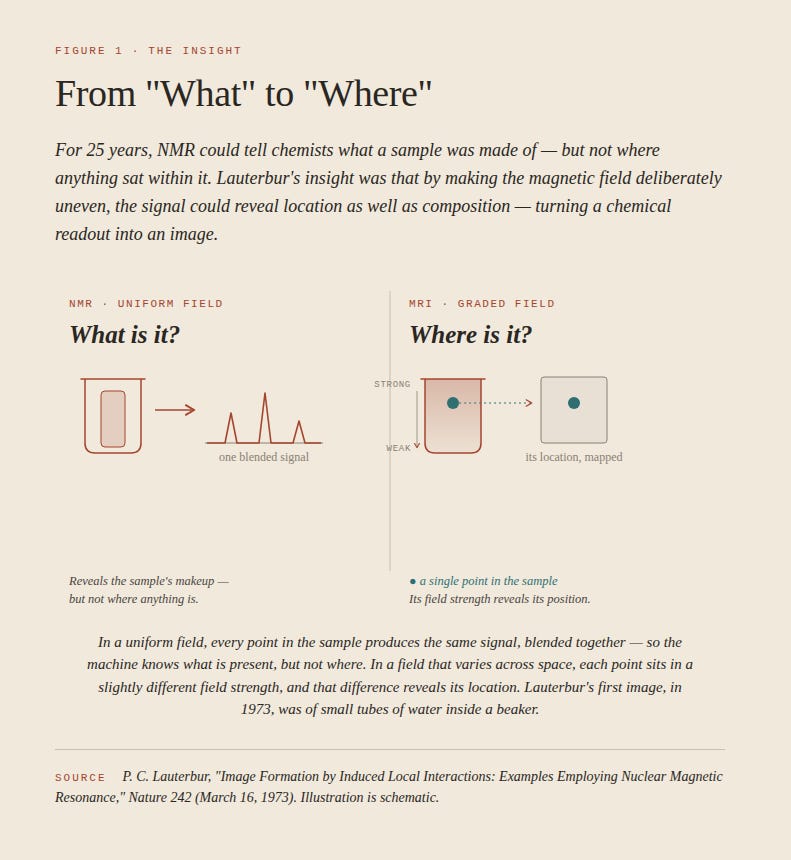
Following this series of experiments, and unsuccessful efforts to patent his idea, he submitted a paper to Nature, describing the results of his work with its images and its implications. The publication initially rejected it — saying Lauterbur’s conclusions were not forceful enough.1 Lauterbur resubmitted the paper with bolder claims, including specific references to medical applications, and the paper was accepted and published on March 16, 1973. At around the same time, Peter Mansfield, a physicist at the UK’s University of Nottingham, was working independently on the same concept. Learning of each other’s work, the two corresponded and influenced each other’s thinking, and in the process Mansfield arrived at a new and much faster method of creating images from the NMR signals. Following this, a number of manufacturers began to develop prototypes that could be used in the field, and by 1980, magnetic resonance imaging (MRI) machines appeared commercially. In 2003, Lauterbur and Mansfield shared the Nobel Prize in Physiology or Medicine for “their discoveries concerning magnetic resonance imaging.”2
An MRI is essentially a giant NMR machine, large enough to hold a person. The human body is roughly 60% water. From an NMR perspective, it is an enormous reservoir of hydrogen atoms which, when placed in a magnetic field and hit with a brief pulse of radio waves, emit a signal — and that signal is what the MRI is measuring. Different tissues contain different amounts of hydrogen. And by deliberately varying the strength of this magnetic field across the body, the scanner causes the hydrogen atoms in different locations in the body to emit signals at slightly different frequencies, allowing the machine to assemble those signals into a map. This was Lauterbur’s key insight. The result is that an MRI scan can create a detailed picture of the body’s soft tissue — for example, the brain, organs, muscles, and joints — which was not possible with other imaging technologies such as X-rays or CT scans. Because an MRI scan uses magnetic fields and radio waves, it introduces no ionizing radiation into the body, and thus can be repeated frequently without risk to the patient. So the Lauterbur insight in 1971 turned a laboratory technique into an imaging tool — and set the stage for a series of specialized applications of the technology, the most consequential of which would arrive in 1990.
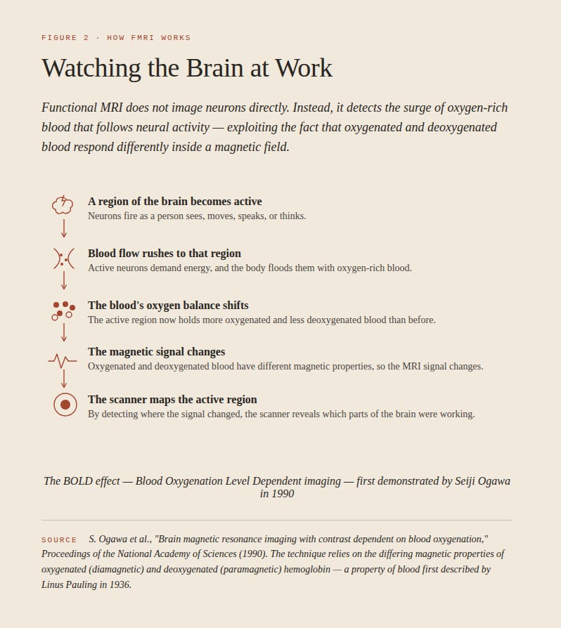
Seiji Ogawa, a Japanese biophysicist working at Bell Labs in New Jersey, had been using MRI to image the brains of mice. Specifically, his 20-year career had been focused on the iron atom and its impact on the magnetic behavior of hemoglobin, the protein in red blood cells that carries oxygen. It had been known since the 1930s that hemoglobin behaves differently, magnetically, depending on the level of oxygen in the blood — this was discovered by Linus Pauling in 1936. What Ogawa noticed in his research was that the MRI images changed depending on the oxygen level in the blood of the mice. It was well established that different areas of the brain become activated during different mental tasks, and that this activation causes changes in blood flow — and therefore changes to blood oxygenation — to that area. Ogawa’s insight was that MRI scans could detect that change, and that, for the first time in history, researchers could watch a living brain — in this case, of a mouse — as it worked. He published his research in December 1990 in the Proceedings of the National Academy of Sciences, naming the phenomenon blood oxygenation level-dependent contrast, or BOLD.3 Through further experimentation, he expanded his work to human brains by 1992, which became the birth of the functional MRI, or fMRI, which would become the dominant tool in the modern study of cognitive neuroscience. This was the beginning of a family of techniques, each based on the imaging capabilities of MRI — which are putting fundamentally new tools for treating diseases in the hands of physicians.
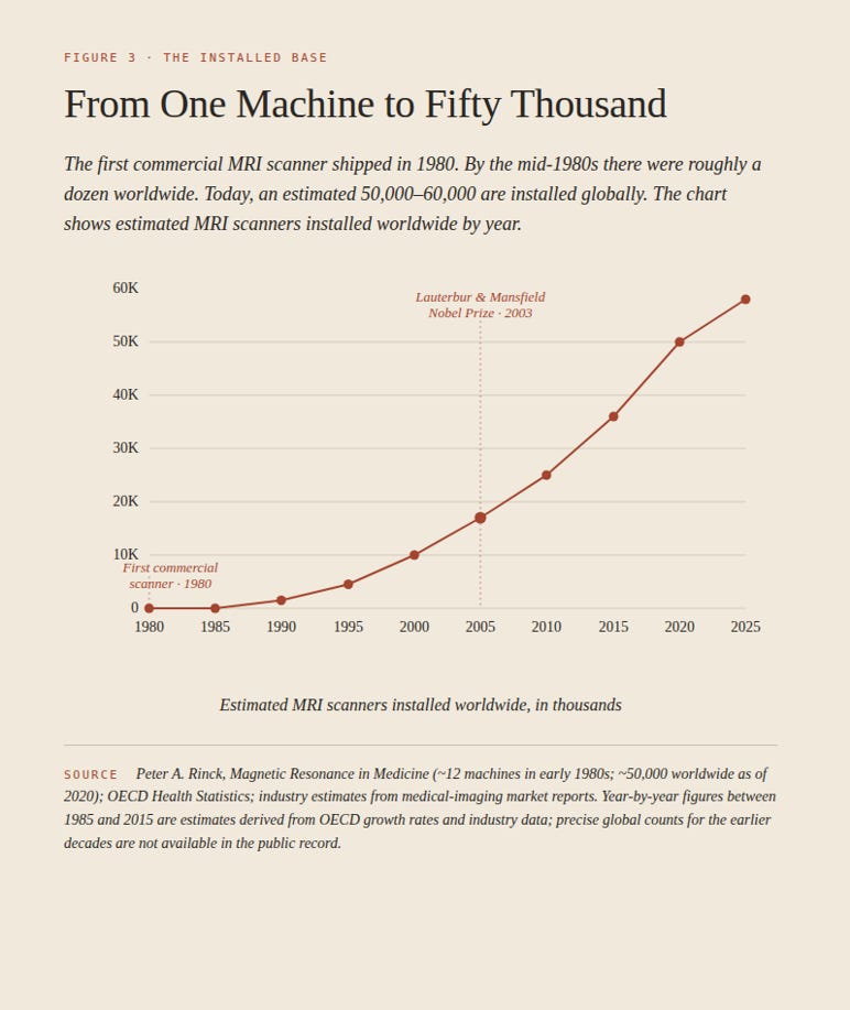
Recent advancements developed along three primary dimensions — reduced cost, faster scans, and more targeted applications. These have increased the deployment of MRI equipment, to the point that there are now 50,000-60,000 machines, and tens of millions of scans performed annually. Meanwhile, the technology continues to gain new powers, from sharper images to shortening scan times — and new applications, such as MRI-guided focused ultrasound, which allows neurosurgeons to destroy small, precisely targeted areas of brain tissue without opening the skull. MRI deployment has also expanded into rural and low-resource settings. The Swoop system, a portable MRI developed by Hyperfine and cleared by the FDA in 2020, costs about $250,000, compared with $1.5 million for a conventional MRI. At the CURE Children’s Hospital of Uganda in Mbale, the Swoop system is now being used to monitor children with hydrocephalus without the radiation of repeated CT scans — a sign this technology is now moving to low- and middle-income countries.4
Neither Lauterbur nor Ogawa developed a new technology. What they did instead was to look at decades-old technologies (Lauterbur) and discoveries (Ogawa), and — drawing on their deep experience in their fields — ask a question that nobody else had thought to ask. Scientists repurpose existing technologies and discoveries all the time — but very few have spread as far as these two have, creating increasingly sophisticated tools to benefit humankind. The question itself is often the key.
References:
1 https://www.thelancet.com/journals/lancet/article/PIIS0140-6736(07)60766-1/fulltext
2 https://www.nobelprize.org/prizes/medicine/2003/summary/